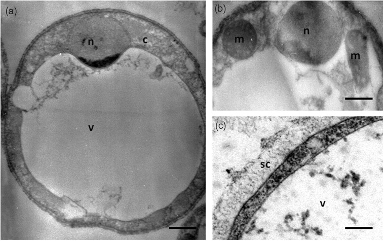
Figure 1.
Vacuolar form of in vitro cultivated Blastocystis sp. viewed under transmission electron microscopy (a). This form is spherical with a large central vacuole (v) and a thin peripheral band of cytoplasm (c) around the vacuole. (b) The cytoplasm contains the nucleus (n) and mitochondrion-like organelles (m). (c) Blastocystis sp. cell is surrounded by a surface coat (sc). Bars, 2 µm for (a) and 500 nm for (b) and (c).

