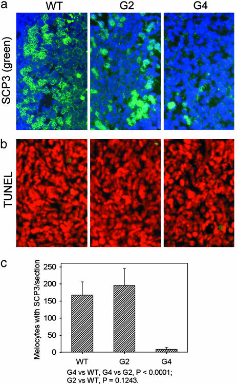
Fig. 2.
Axial/lateral elements of synaptonemal complexes and apoptosis in WT, G2, and G4 TR-/- fetal ovaries. (a) Meiocytes with SCP3 elements (green) and nuclear chromosomes (blue). (b) Very few apoptotic cells (green) in WT, G2, and G4 TR-/- fetal ovaries. Red, nuclear staining with propidium iodide. (c) Number of meiocytes with SCP3 elements per section of fetal ovaries, showing significantly reduced SCP3 meiocytes in G4 TR-/- fetal ovaries, compared with WT and G2 TR-/- fetal ovaries.

