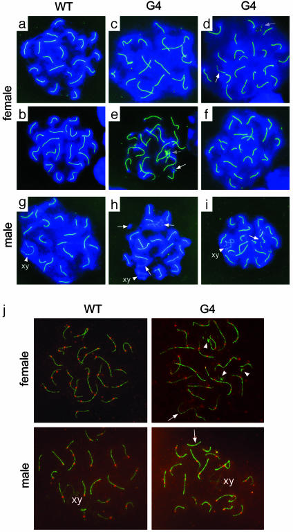
Fig. 4.
Immunostaining of SCP3 elements and MLH1 foci at pachytene stage from female fetal ovary (day 17.5 of gestation) and male testis (day 20 after birth). (a–i) SCP3 elements and structure. WT pachytene oocytes show 20 SCP3 elements (a and b), whereas G4 TR-/- pachytene oocytes show 19 (c), 21 (d), 22 (e), and 21 (f) SCP3 elements. WT pachytene spermatocytes show 20 SCP3 elements (g), but G4 T-/- pachytene spermatocytes show altered number of SCP3 elements (21) (h and i). Green, SCP3; blue, chromosomes counterstained with DAPI; white arrows, unpaired lateral SCP3; gray arrows, SCP3 fragments. (j) MLH1 foci and SCP3 elements. Only those MLH1 dots (red) unambiguously colocated with SCs (green) are counted as MLH1 foci crossover sites (27, 28). Some MLH1 foci are present on the diffuse chromatin surrounding SCs and are considered as MLH1 background staining (27). Arrows, lack of MLH1 foci in the SCP3 elements; arrowheads, SCP3 fragments.

