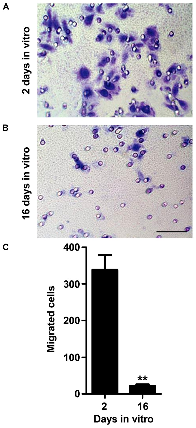
FIGURE 3.
Microglia migration ability decreases with cell aging in culture. Microglial cells were kept in culture for 2 and 16 days in vitro (DIV) and then cellular chemotactic migration to 10 μM ATP was evaluated using the Boyden chamber method. Representative images of 2 (A) and 16 (B) DIV microglia that migrated towards ATP were visualized by Giemsa staining. Number of migrated cells was counted and results expressed in graph bars as mean ± SEM (C). Cultures, n = 4 per group. t-test, **p < 0.01 vs. 2 DIV cells. Scale bar equals 50 μm.

