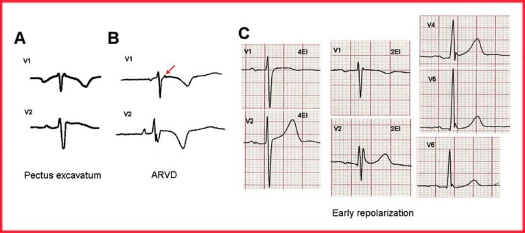
Fig. (5).
(A) The ECG pattern of Pectus excavatum (22) depicting a negative P-wave in lead V1 (electrodes in adequate position). The r'-wave in lead V1 is narrow, followed by a slight ST-segment elevation. (B) In ARVD, the ECG pattern is quite different from that of BrS. The presence of an epsilon wave (arrow), atypical RBBB and negative T-waves beyond lead V3 helps in distinguishing between these two entities. (C) The ECG of patients with early repolarization usually presents J point and ST-segment elevation in the middle and left precordial leads. However, if leads V1-V2 electrodes are placed in a non-standard position (2nd intercostal space), rSr’ morphology is observed.

