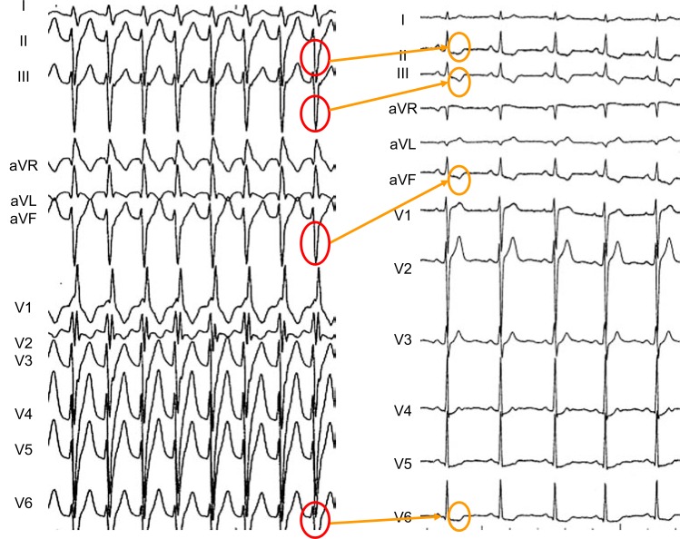
Fig. (3).
Fascicular ventricular tachycardia (left) showing a pattern of “right bundle branch block and left anterior hemiblock”. The wide QRS are predominantly negative in leads II, III, aVF and V3 to V6. After spontaneous termination of the arrhythmia, abnormal “memory-.induced” T waves are observed in leads II, III, aVF and V4 to V6. Note that post- tachycardia T waves are positive in leads V1 and V2, in which the conditioning QRS complex was also positive. This is an example of short-term T wave memory, appearing after a short period of abnormal ventricular depolarization.

