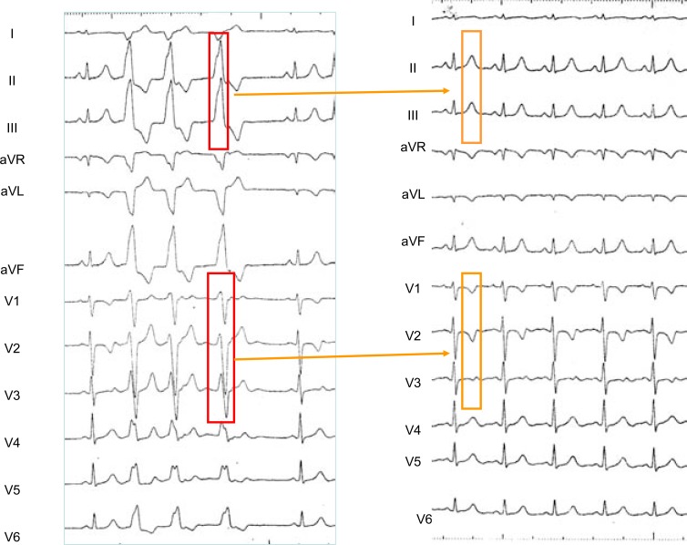
Fig. (7).
Left panel: 12 simultaneous ECG leads obtained in a patient with incessant self-limited episodes of ventricular tachycardia with a “LBBB like” pattern before ablation of the ectopic focus). Ventricular ectopy shows tall R waves in the inferior leads and its precordial transition is situated in lead V3. Right Panel: ECG recorded after ablation of the focus found situated in the left aortic sinus of Valsalva, 1cm below the left coronary artery ostium. The “memory-induced” T waves spatial orientation exactly reproduces that of the QRS complexes of ventricular tachycardia.

