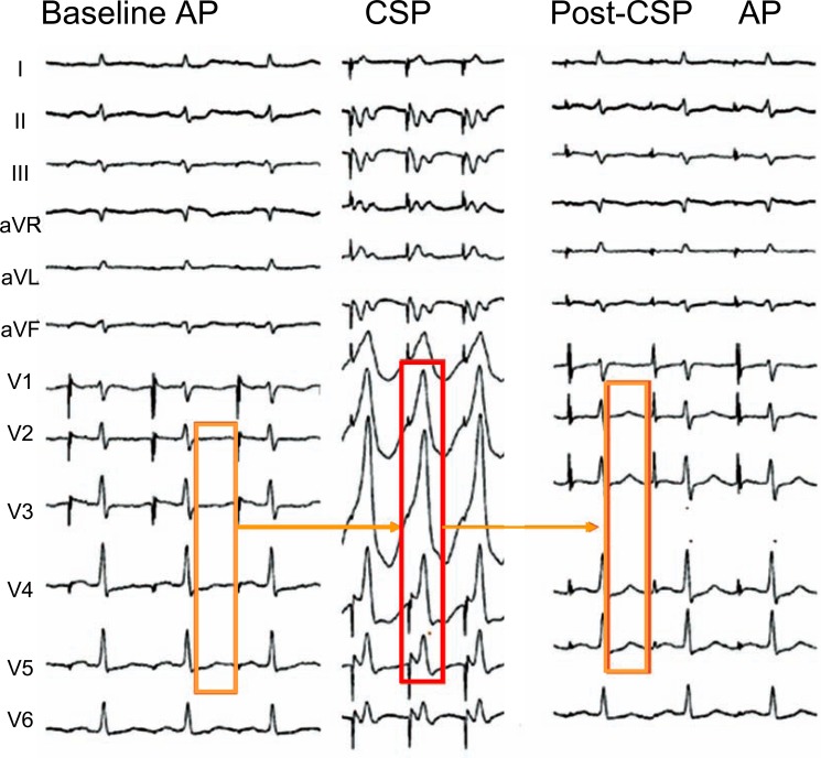
Fig. (9).
Memory-induced normalization of primary abnormal T waves in precordial leads in a 57 year old patient with idiopathic dilated cardiomyopathy and syncope. The baseline ECG recorded during atrial pacing (AP) at a cycle length (CL) of 750 ms showed abnormal negative or flat T waves in leads V1 to V6 (A). After a 15-minute period of left ventricular pacing (VP) at a CL of 500 ms (B), the T wave became less negative in lead V1 and ”normalized” in V2-V6, tracking the predominant electrical forces of the previously paced QRS complexes (C).

