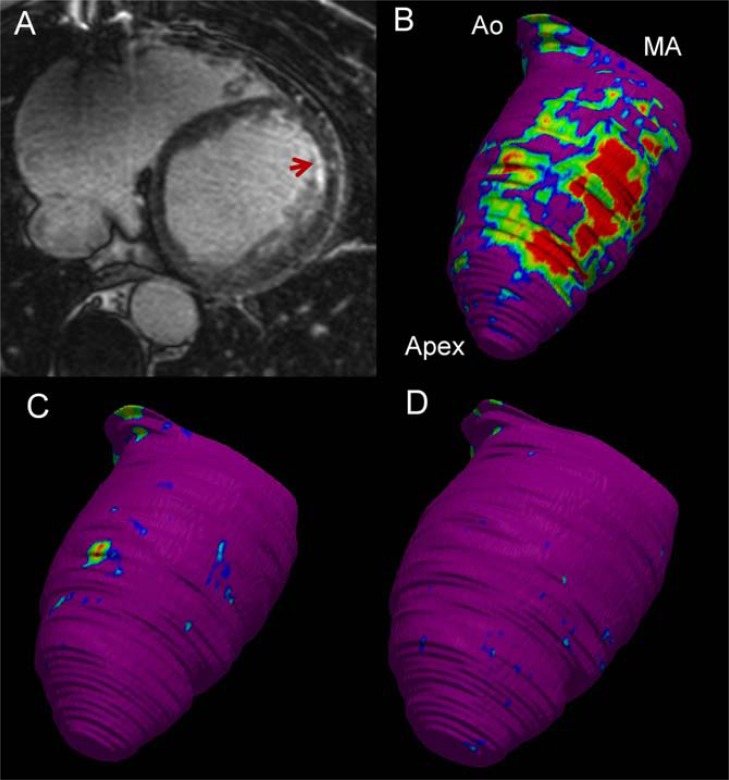
Fig. (5).
Contrast-enhanced cardiac magnetic resonance (CeCMR) from a patient with subendocardial anterior myocardial infarction and ventricular tachycardia (12-lead electrocardiogram is shown in the right panel of Fig. (3)). Panel A shows axial-axis view of CeCMR showing subendocardial hyperenhancement in the anterior wall of the left ventricle. Panels B to D show signal intensity maps obtained from CeCMR projected over 3D color-coded shells at subendocardium (panel B), midmyocardium (panel C) and subepicardium (panel D). Normal myocardium is coded in purple, core of the infarct in red and border zone in blue-green-yellow. Identification by CeCMR of scar confined to subendocardium indicates that an endocardial approach will be adequate to abolish the arrhythmia. Apex left ventricular apex; Ao, Aortic root; MA, mitral annulus.

