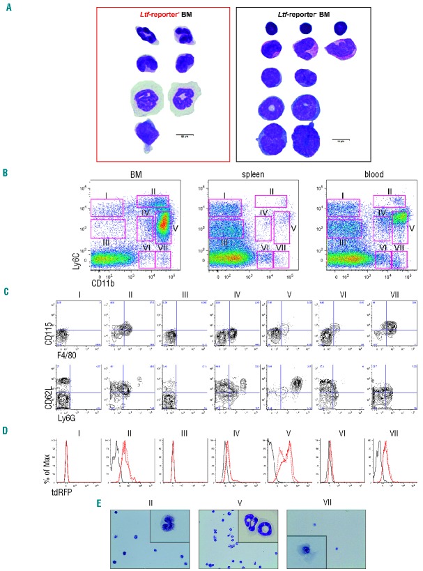
Figure 5.
Ltf-reporter expression in monocytes, macrophages and neutrophils. (A) Morphology of Ltf-reporter+ and Ltf-reporter− BM cells was determined by Grünwald-Giemsa staining from sorted fractions. Representative cells from individual images of the same specimen were grouped with regard to their morphology as described elsewhere.24 Notably, eosinophils were enriched specifically in the Ltf-reporter− BM fraction. (B–E) Murine BM, spleen and peripheral blood cells from Ltf-reporter+ mice were stained with antibodies against CD11b, Ly6C, Ly6G, CD115, F4/80 and CD62L. (B) Resolution of live cells with CD11b and Ly6C reveals seven distinct populations (I–VII). (C) Each population was further gated for CD115 and F4/80 (upper panels) and CD62L and Ly6G (lower panels). (D) Histograms depict the intensity of Ltf-reporter in each of the seven populations. Populations II, V and VII consist of Ltf-reporter+ cells. (E) Morphology of populations II, V and VII was determined by Grünwald-Giemsa staining on cytospin preparations from sorted cells. Ly6Chigh/CD11b+/CD115+/F4/80+/CD62L+/Ly6Glow cells resemble monocytes (population II), while Ly6C+/CD11bhigh/CD115−/F4/80−/CD62L+/Ly6Ghigh cells consist of ring-shaped neutrophilic precursors. Ly6C−/CD11bhigh/CD115+/F4/80+/CD62Llow/Ly6Glow resemble macrophages.

