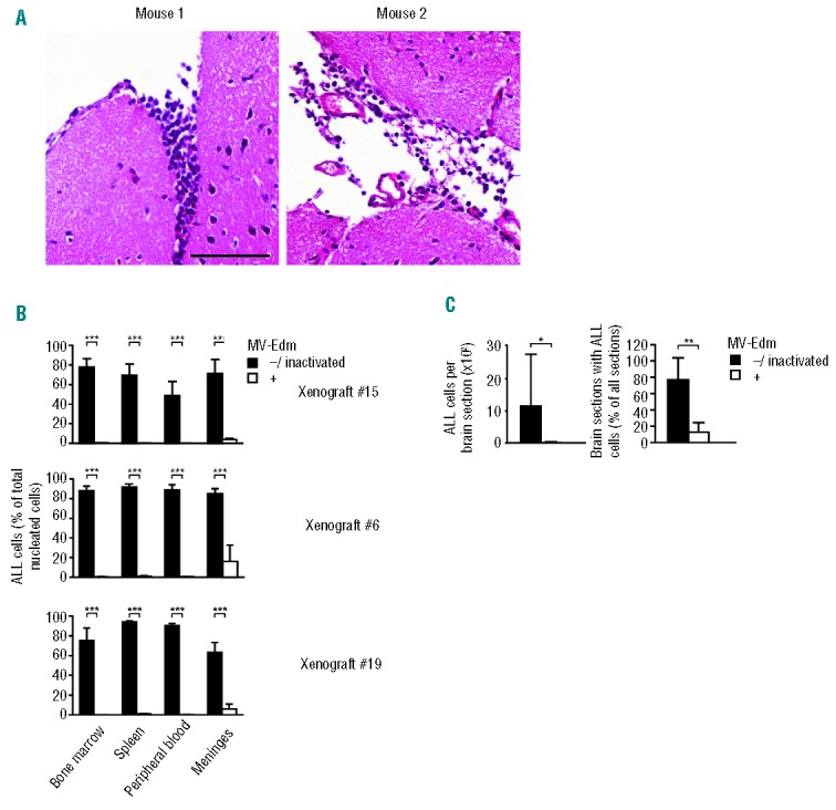
Figure 7.

Systemic MV-Edm curbs CNS disease In NOD/SCID mice with ALL xenografts (A) CNS disease is established early. 2 mice with xenograft #21 were killed once peripheral leukemic blasts reached 5–20 % (90 d), as determined by FACS, and CNS disease was assessed by FACS analysis of the meninges and histology of the brain. Scale bars equal 100 μm. (B) Meningeal blasts are controlled while not eradicated by MV-Edm. Mice with xenografts without MV-Edm (“−” and “inactivated”) that thus died from disease, and which could be assessed before onset of autolysis (8 out of 16 mice for xenograft #15, 7 out of 8 for xenograft #6, 10 out of 10 for xenograft #19 and 9 out of 10 for xenograft #13) were compared to mice that were treated with MV-Edm (“+”) and thus died or were sacrificed because of advanced age (8 out of 8 mice for xenograft #15, 7 out of 9 for xenograft #6, 8 out of 9 for xenograft #19 and 2 out of 10 for xenograft #13). Organs were investigated by FACS for the presence of CD45+Ly5− leukemic cells. ***P<0.001 using the unpaired t-test. (C) Marked decrease of blasts infiltrating the brain. Brains of mice with xenograft #15 were serially cut as depicted in Online Supplementary Figure S7 and stained by HE. Blasts were detected by morphology (left panel, scale bar equals 100 μm). The number of leukemic cells per section and the percentage of sections containing leukemic cells were determined (right panel; means +SD for each group are depicted; **P<0.01, *P<0.05 using the unpaired t-test).
