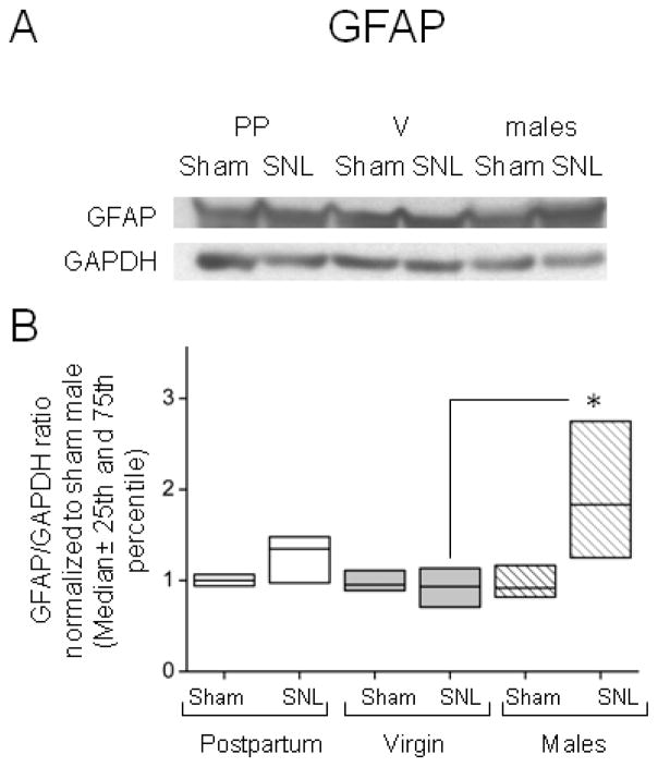
Figure 3.
Sexual dimorphic differences in the GFAP expression after SNL surgery determined by western blot analysis. (A) Representative immunoblot for GFAP and GAPDH from ipsilateral dorsal lumbar spinal cord homogenizates. (B) The expression of GFAP increases after SNL surgery in male rats (stripes) when compared to virgin females rats (V (light grey)). Data represent the GFAP/GAPDH ratio, normalized to the median value for the Sham male group. Data are presented as median (horizontal line) ± 25th and 75th percentiles; n=7 in each group of females and n=5 in each group of males, Kruskal-Wallis one way analysis of Variance on Ranks (p=0.029), using Pairwise multiple comparison procedures (Dunn’s method), *p <0.05 compared to VSNL.

