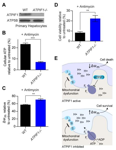
Figure 4. Inhibition of ATPIF1 ameliorates the effects of complex III blockade in primary hepatocytes.
(A) Immunoblots for indicated proteins of primary hepatocytes derived from WT and ATPIF1 -/- mice. (B) Cellular ATP of WT and ATPIF1 -/- primary hepatocytes treated with antimycin (0.625 μM) for 1.5 hours. Error bars are ± s.e.m. (n = 3). ***P < 0.001. (C) ΔΨm of WT and ATPIF1 -/- primary hepatocytes treated with antimycin (10 μM) for 1.5 hours. Error bars are ± s.e.m. (n = 3). **P < 0.01. (D) Viability of WT and ATPIF1 -/- primary hepatocytes treated with antimycin (1.25 μM) for 2 days. Error bars are ± s.e.m. (n = 3). **P < 0.01. (E) Schematic diagramming the behavior of cells with ETC dysfunction under conditions where ATPIF1 is active or inhibited. Inhibition of ATPIF1 is depicted by absence of the protein but represents any strategy to block ATPIF1 activity on the F1-F0 ATP synthase. See also Figure S4.

