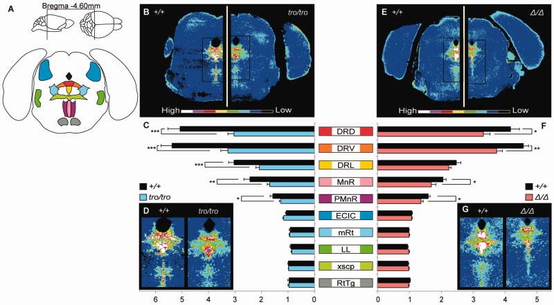
Fig. 6.—
Quantitative immunohistochemistry for TPH2. (A) Fluorescence intensities were measured in transverse sections located approximately 4.60 mm caudal from bregma, including 5-HT-rich region, the raphe nucleus. Abbreviations (Franklin and Paxinos 2008): DRD, dorsal part of the dorsal raphe nucleus; DRV, ventral part of the dorsal raphe nucleus; DRL, lateral part of the dorsal raphe nucleus; MnR, median raphe nucleus; PMnR, paramedian raphe nucleus; ECIC, external cortex of the inferior colliculus; mRt, mesencephalic reticular formation; LL, lateral lemniscus; xscp, decussation of the superior cerebellar peduncle; RtTg, reticulotegmental nucleus of the pons. (B–D) Quantitative distribution, comparative analysis, and enlarged the raphe nucleus for +/+ and tro/tro, respectively. (E–G) Quantitative distribution, comparative analysis, and enlarged the raphe nucleus for +/+ and Δ/Δ, respectively. Values shown on graphs represent the mean ± SEM. *P < 0.05; **P < 0.01; ***P < 0.001.

