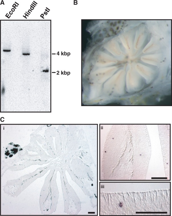
Fig. 1.—

V1R2 copy number and mRNA expression within the olfactory epithelium. (A) Southern blot analysis of genomic DNA isolated from Haplochromis chilotes (heterozygote of V1 and V9) using a V1R2 probe. DNA was digested with EcoRI, HindIII, or PstI before electrophoresis. Size markers are indicated to the right. Photographs from two independent experiments were combined. (B) The photo of the olfactory organ (called “olfactory rosette”) of the adult individual of H. sauvagei. (C) Three magnifications of the thin sections of the olfactory rosette are shown. Horizontal sections (7 µm) were hybridized with a DIG-labeled cRNA probe to evaluate V1R2 (V9 allele) expression. Scale bars indicate 100 µm.
