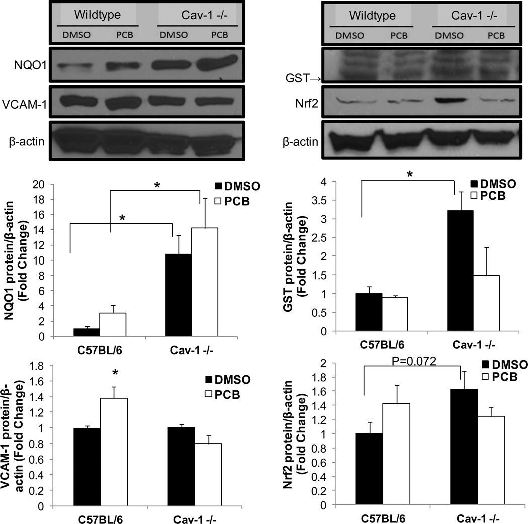
Fig. 5.
Confirmation of Cav-1 Nrf2 cross-talk in cells isolated from Cav-1 −/− mice. Endothelial cells from Cav-1 −/− and age matched wildtype control mice were isolated and subsequently exposed to PCB 126 at a concentration of 2.5 µM for 24 h. Protein levels of NQO1, VCAM-1, GST and Nrf2 were measured via Western blot. Results were normalized to β-actin and depicted as fold change compared to control siRNA DMSO treatments. Results represent the mean±SEM (NQO1, GST, Nrf2: n=3, VCAM-1: n=2 for each treatment group). Cells isolated from Cav-1 −/− mice did not display PCB-induced VCAM-1 upregulation and showed significantly increased levels of NQO1 (*p<0.05). Cells isolated from Cav-1 −/− mice displayed significantly higher levels of GST under DMSO vehicle conditions compared to wildtype cells treated with DMSO (*p<0.05). Also, cells isolated from Cav-1 −/− mice displayed a trend toward being significantly increased compared to vehicle treated control cells. Western blots above show representative samples of visual results.

