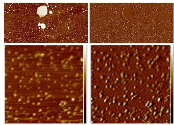
Figure 4.
After extended incubation in buffer, lipin adheres strongly enough to the mica to be visualized. An area of bare mica adjacent to the patch of bilayer seen in Figure 3 is shown at low magnification in the top panels (2.4 × 5 μm), left panel height image, right panel amplitude error image of the same region. The large bright objects in the height image are small patches of lipid bilayer, the small particles on the mica are lipin. The lower panels show a portion of the same field at higher magnification (750 nm × 750 nm), left panel height image, height scale −2 to + 2 nm, right panel corresponding amplitude error image, amplitude error scale −10 to +10 mv. The lipin particles move slightly toward the upper right under the force of the probe, distorting the appearance of the particles.

