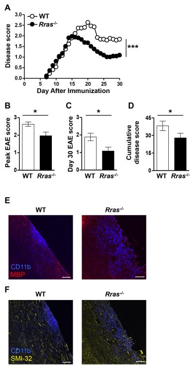
Figure 1. Rras−/− mice exhibit attenuated EAE disease severity.
A-F, EAE was induced in WT or Rras−/− mice at 6-8 wk of age by s.c. immunization with MOG35-55 peptide. A, Clinical signs of EAE were evaluated daily starting on day seven and the data shown are the daily average of 15 WT and 15 Rras−/− mice. B, The mean ± SE peak disease score is shown for WT mice on day 20 and day 15 for Rras−/− mice. C, The mean ± SE disease score at the end of the experiment on day 30 is shown. D, The cumulative disease score was calculated by adding the daily scores of each mouse from day 7-30 and shown as the mean ± SE of all the mice in each group. E-F, longitudinal frozen sections from the spinal cord of mice 17 days after EAE induction were generated from WT and Rras−/− mice. The sections were stained with anti-CD11b (blue) (E,F), anti-MBP (red) (E) and with the SMI-32 antibody (yellow) (F) and analyzed by immunofluorescence. Overlaid images of CD11b and MBP (E) or SMI-32 (F) are shown. Data shown are representative of two mice in each group. *p<0.05, ***p<0.001. Scale bar: 80 μm.

