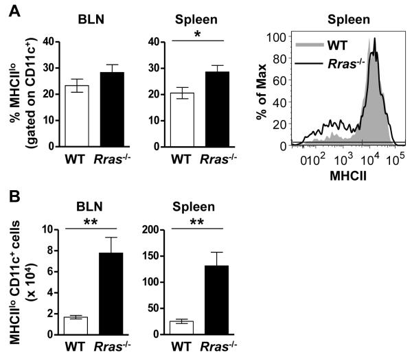
Figure 4. Increased numbers of MHCIIlo DC are present in the BLN and spleen of Rras−/− mice during EAE.
A,B, EAE was induced as for Fig. 1 and the presence of MHCIIlo DC was evaluated 17 days later in WT and Rras−/− mice. A, The percentage of CD11c-gated MHCIIlo cells in the BLN (left panel) and spleen (middle panel) is shown. One representative experiment showing MHCII expression levels in the spleen is shown (right panel). B, The absolute number of CD11c-gated MHCIIlo cells in the BLN (left panel) and spleen (middle panel) is shown. Data shown are the mean ± SE of two independent experiments with six mice in each group. *p<0.05, **p<0.01.

