
Figure 2. TSLP receptor components are expressed in sensory neurons.
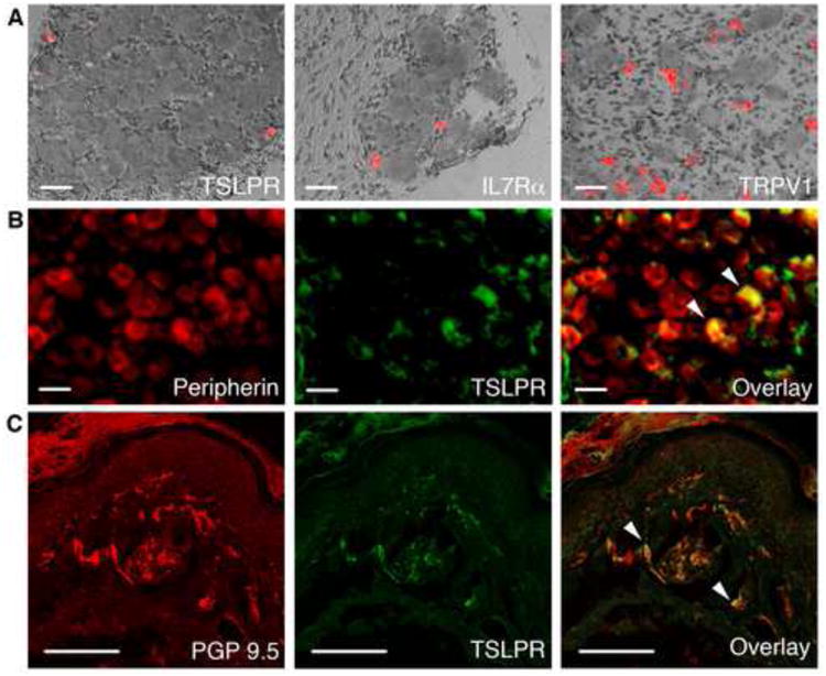
(A) DIC overlay images of in situ hybridization with cDNA probes detecting TSLPR, IL7Rα and TRPV1 in mouse DRG. Scale bar = 400μm. (B) Immunostaining of DRG sections with antibodies against peripherin and TSLPR in DRG sections. White arrows (right) mark peripherin- and TSLPR-positive neurons. Scale bar = 400μm. n≥4 mice/condition. (C) Immunostaining of PGP 9.5 and TSLPR in glabrous hind paw skin. The white arrows (right) mark PGP 9.5- and TSLPR-positive neurons. Scale bar = 200μm. n≥3 mice per condition.