
Figure 3. TSLP directly activates a subset of sensory neurons.
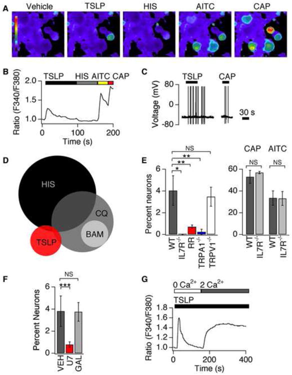
(A) Representative images of Fura-2 loaded DRG neurons treated with vehicle, TSLP (2 ng/mL), histamine (HIS, 1mM), AITC (200μM) and capsaicin (CAP, 1μM). (B) Representative trace shows a neuron that responds to TSLP, AITC and CAP, but not HIS. (C) Current-clamp recording showing TSLP- and CAP-evoked action potential firing in a DRG neuron. n≥60 cells. (D) A small percentage of the TSLP-sensitive population overlaps with the population of histamine- (HIS, 6%) or chloroquine-sensitive neurons (CQ, 6%), but not the BAM8-22 population (BAM, 0%). (E) Left: Prevalence of TSLP sensitivity in wild-type neurons (black), IL7Rα-deficient (grey) neurons, neurons treated with 20μM ruthenium red (RR; red), TRPA1-deficient neurons (blue) and TRPV1-deficient neurons (white). Right: prevalence of AITC and CAP sensitivity in wild-type (black) and IL7Rα-deficient (grey) neurons n≥1000 cells. (F) Prevalence of TSLP sensitivity in neurons pre-treated with vehicle (black), a PLC blocker, U73122 (red) and the Gβγ blocker, gallein (grey) n≥600 cells. (G) Representative response to TSLP in the absence (0mM Ca2+) and presence (2mM Ca2+) of extracellular Ca2+ n≥200 cells. *P<0.05; **P<0.01; ***P<0.001. Error bars represent s.e.m.