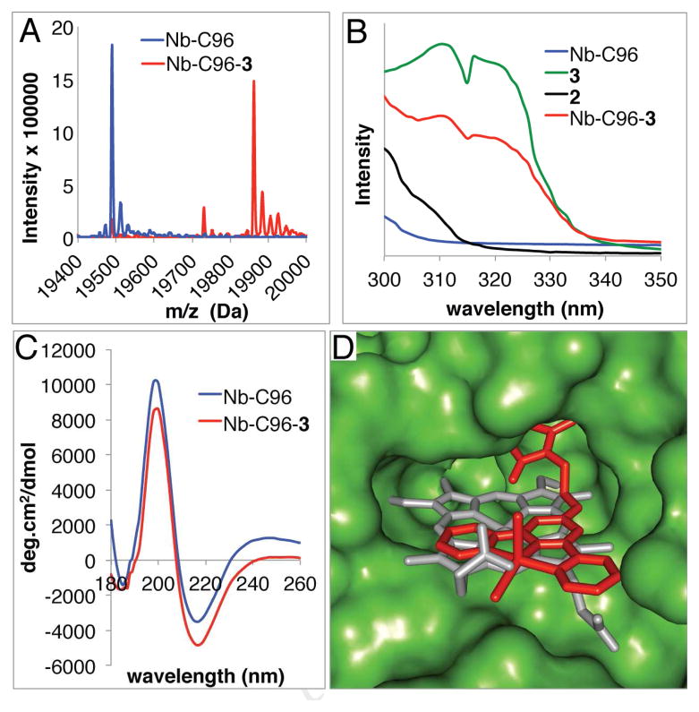
Figure 1.
A) HR ESI-MS spectra of Nb-C96 and Nb-C96-3 showing the efficiency of Nb bioconjugation. B) UV spectra of Nb-C96, Nb-C96-3, 2, and 3 showing the presence of MnCl2 in Nb-C96-3. C) CD spectra of Nb-C96 and Nb-C96-3 showing proper Nb folding following bioconjugation. D) Overlay of a DFT-optimized structure of 3 (red) covalently linked (Pymol) to a crystal structure of Nb (PDB ID 3EMM27) and the native heme (gray).

