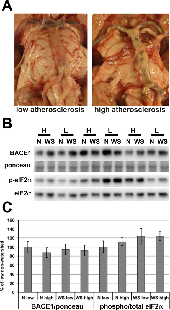
Fig. 1. BACE1 and phospho-eIF2α levels are not elevated in non-demented individuals with high cerebrovascular disease.
(A) Representative images of the circle of Willis from non-demented individuals categorized as having low or high levels of atherosclerosis. (B) Homogenates of human brain samples described in (Table 1) were analyzed by immunoblotting for BACE1 and phospho- and total eIF2α, as shown in representative blots. (C) Quantification of immunoblots shows no difference in BACE1 or ratio of phospho to total eIF2α in those with high levels of atherosclerosis compared to those with low atherosclerosis. The blot in (B) shows only 6 individuals in the interest of space. This is just a portion of the blot containing both watershed and non-watershed regions from all 19 individuals that was used in quantification. N=non-watershed, WS= watershed, H=high atherosclerosis, L=low atherosclerosis. p<0.05 *

