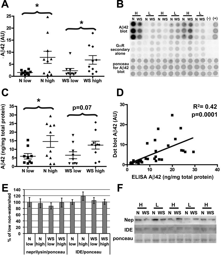
Fig. 2. Aβ42 levels are elevated in non-demented individuals with atherosclerosis, but neprilysin and insulin degrading enzyme levels are unchanged.
(A, B) Brain homogenates extracted in guanidine hydrochloride were analyzed by Aβ42-specific dot blot and quantified. (C, D). The accuracy of the Aβ42 dot blot was verified by subjecting the same samples to a commercial Aβ42-specific ELISA. The two methods yielded similar results. In addition, there is significant correlation between the level of Aβ42 measured by ELISA and dot blot (D). Combined, these data show an elevation in Aβ42 in the high atherosclerosis group in both watershed and non-watershed regions, and no difference between the regions in either low or high atherosclerosis groups. (E, F) Analysis by immunoblot and quantification shows that the increase in Aβ42 cannot be explained by a decrease in levels in either of the Aβ degrading enzymes, neprilysin or IDE as there is no difference between high and low atherosclerosis groups in either region. As in (Fig. 1), (B) and (F) show only 6 and 5 representative individuals respectively, in the interest of space, but these are just portions of the blots containing both watershed and non-watershed regions from all 19 individuals that were used in our analysis. N=non-watershed, WS= watershed, H=high atherosclerosis, L=low atherosclerosis.

