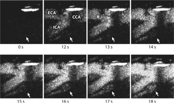
Figure 1.
Contrast-enhanced ultrasound of the carotid plaque. Time 0 s shows the total subtracted image (black) in the nonlinear imaging mode at the carotid bifurcation before the arrival of the contrast agent. From time 12 s, the enhancement of the carotid lumen (white) is evident. Note the subsequent enhancement of the plaque neovascularization from time 14 s (white arrow). CCA, common carotid artery; ECA, external carotid artery; ICA, internal carotid artery. Reproduced with permission from Prof. Ed Leen, Imperial College London.

