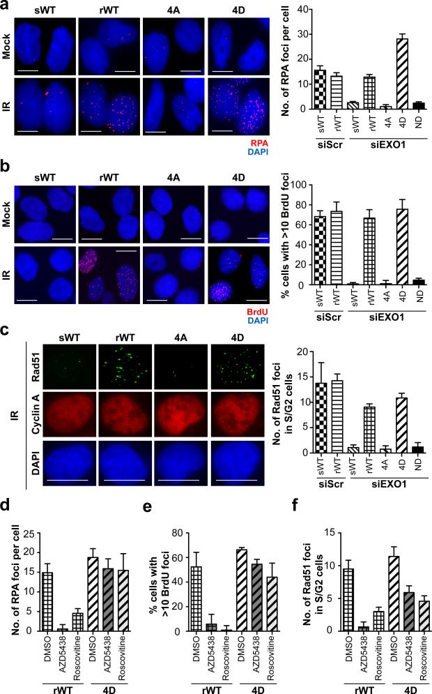
Figure 2. Phosphorylation of EXO1 by CDKs promotes DNA end resection.
(a) U2OS cells were depleted of endogenous EXO1 using siRNA and complemented with siRNA-sensitive wild type EXO1 (sWT) or siRNA-resistant wild type EXO1 (rWT), nonphosphorylatable mutant EXO1 (4A), or phospho-mimic EXO1 with all four C-terminal S/TP sites replaced with Asp (4D), as indicated. Representative images of mock-irradiated or irradiated (IR) cells immunostained with anti-RPA antibody (red) are shown. Nuclei were stained with DAPI (blue). Plot shows average numbers of RPA foci per cell after subtracting background (average numbers of foci in mock-irradiated cells). (b) Representative images of mock-irradiated or irradiated cells immunostained for BrdU/ssDNA foci (red) are shown. Plot shows percentages of cells with more than 10 BrdU foci per nucleus after background subtraction. (c) Representative images of irradiated cells co-immunofluorescence stained with anti-Cyclin A antibody (red) and anti-Rad51 antibody (green) are shown. Average numbers of Rad51 foci for Cyclin A positive (S/G2) nuclei are plotted after background subtraction. (d,e,f) Cells expressing rWT or 4D EXO1 were pre-treated with CDK inhibitors (AZD5438 or Roscovitine) or with DMSO as control, irradiated, and assayed for RPA, BrdU, or Rad51 foci, as indicated. Scale bars denote 10 μm for all images. All experiments were replicated three times. Error bars depict S.E.M. See also Supplementary Fig. 2.

