Fig. 3.
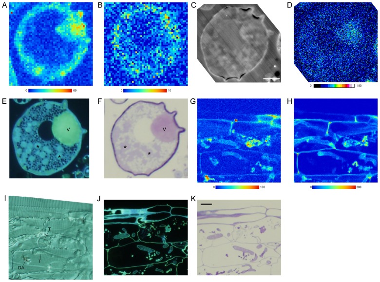
Plunge-frozen auxiliary cells of Gigaspora margarita produced on an extraradical hypha (A–F) and mycorrhizal roots (G–K) of Allium cepa mycorrhiza exposed to Cd solution. A, B, G, & H, Synchrotron radiation micro X-ray fluorescence (SR-μXRF) imaging of Cd (A, G), and Zn (B, H) (step, 1 μm; exposure time, 1 sec). Color bars indicate minimum and maximum X-ray counts. C & D. SEM-COMPO images of the section used for SR-μXRF (C) and phosphorus (Kα, 3.3 keV) mapping by EDS-SEM (D). Color bar indicates x-ray counts described by gray values. E & J, 4′,6-diamidino-2-phenylindole staining for polyphosphate of a serial semi-thin section observed by a fluorescence microscope. F & K, Toluidine blue O staining of a serial semi-thin section viewed by a light microscope. I, A differential interference image of a 10 μm thick section used for SR-μXRF. Asterisk, nucleus. DA, dead arbuscule; T, trunk hypha; V, vacuole. Bar: C, 10 μm; K, 20 μm.

