Figure 6.
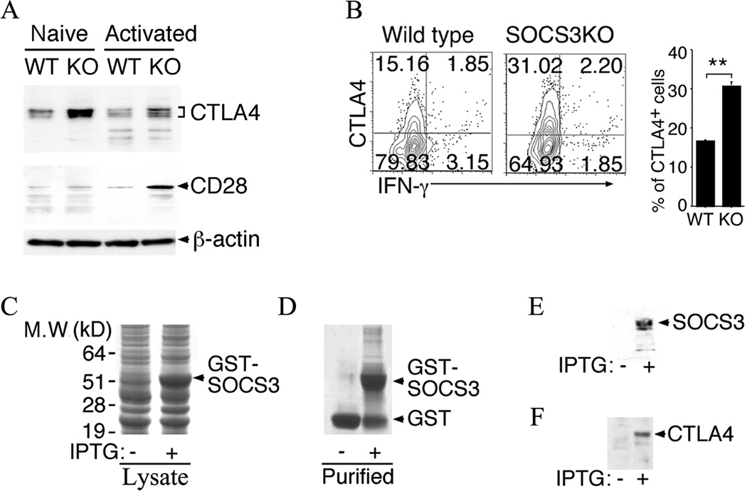
SOCS3 interacts with CTLA-4 and down-regulates its expression in CD4+ T cells. (A) Naïve CD4+ T cells were stimulated by anti-CD3/CD28 for three days and analyzed by Western blotting. (B) Freshly isolated CD4+ T cells isolated from IRBP-immunized mice were analyzed by the intracellular cytokine staining assays. The cells were gated on CD4 and numbers in quadrants indicate percent of CD4+ T cells expressing CTLA-4 and/or IFN-γ. (C–F) A GST-SOCS3 fusion protein was produced by cloning the full-length mouse SOCS3 cDNA into the pGEX6p1 expression vector and analyzed by SDS-PAGE, western blotting and/or immunoprecipitation. (C) Crude extracts (20µg) from transformed E. coli BL21 bacterial GST-SOCS3 recombinants bacteria before and after induction with IPTG were fractionated SDS-PAGE and stained with Coomassie blue dye. (D, E) Affinity purified GST-SOCS3 protein was analyzed by SDS-PAGE/Coomassie blue staining (D) or Western blotting using anti-SOCS3 Abs (E). (F) Western blot analysis following pull-down of CTLA-4 from activated CD4+ T cell extracts by GST-SOCS3. Results are representative of at least three independent experiments.

