Fig. 7.
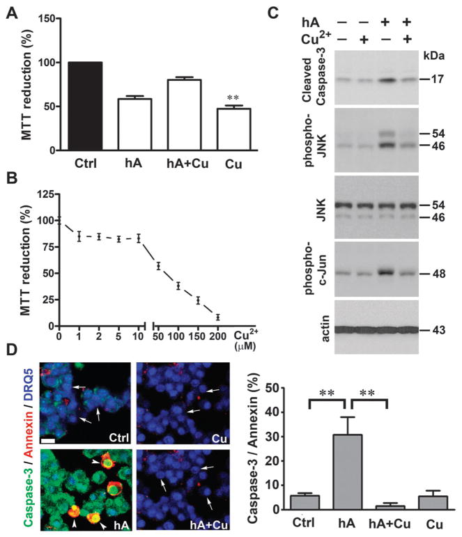
Cu2+-inhibits amylin-evoked apoptosis in pancreatic cells. Human amylin (20 μM) was incubated in the absence or presence of 10 μM CuCl2 and corresponding changes on cells’ viability were determined using MTT reduction (A and B) and apoptotic western-blot (C) and microscopy (D) assays. (A) Amylin induced a marked drop in the MTT signal, indicative of metabolic stress in cells. (B) Cu2+ impaired mitochondrial enzymatic activity in a dose-dependent manner. (C) Western blot analysis reveals an inhibitory effect of Cu2+ on caspase-3 and stress kinases activity in cells treated with human amylin. (D) Confocal microscopy analysis of apoptosis in RIN-m5F cells treated with human amylin, in the absence and in the presence of Cu2+. Arrows depict non-apoptotic nuclei (blue) in viable cells, whereas arrowheads depict caspase-3 (green) and annexin-V (red) positive apoptotic cells. Bar, 10 μm. Quantitative analysis of amylin-induced apoptosis in human islets is summarized in a bar graph. Significance established at **P < 0.01, n = 6, ANOVA followed by Newman–Keul post hoc test.

