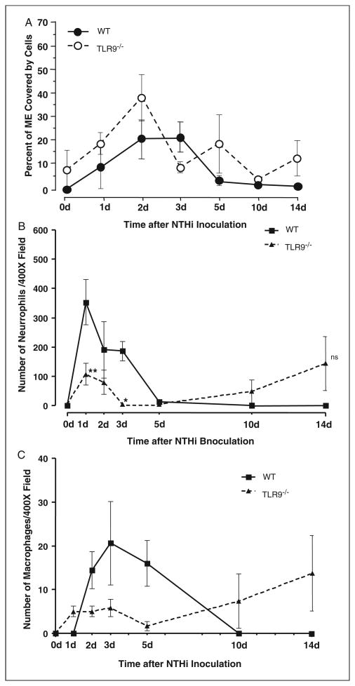
Figure 4.
Leukocyte infiltration of the ME cavity in wild-type versus TLR9−/− mice. (A) Inflammatory cell infiltration into the ME lumen of wild-type and TLR9−/− mice during OM, measured as a percentage of ME luminal area covered by cells. There was no significant difference in the recruitment of cells to the MEs of TLR9−/− mice during OM. n = 6–8 MEs per time point; bars represent the mean ± SEM. (B) Neutrophils increased significantly less from days 1-3 compared to wild-type mice, but their presence in the ME was prolonged. (C) There was no significant difference in the recruitment of macrophages to the TLR9−/− ME. n = 6–8 MEs per time point; bars represent ±SEM.

