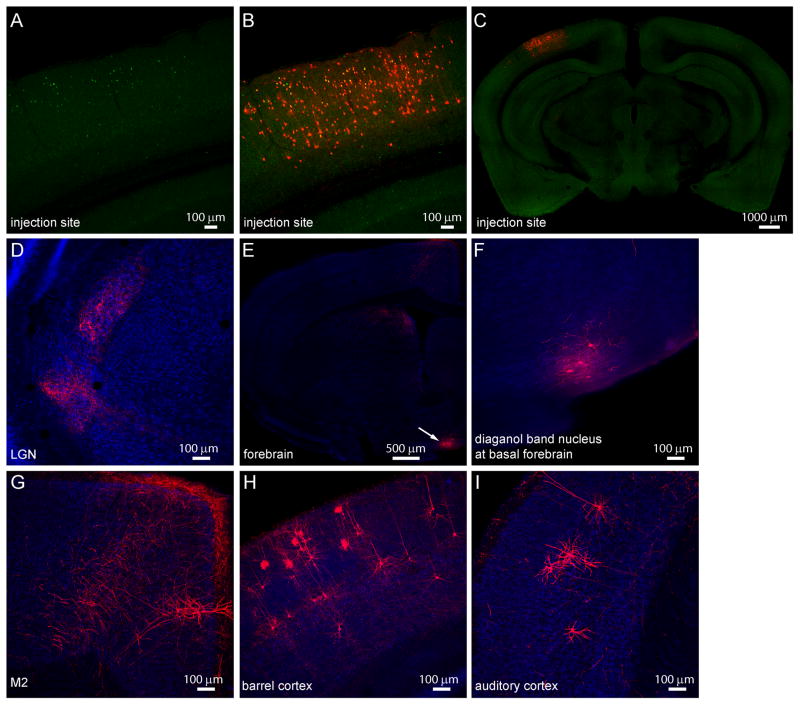
Figure 5.
Retrograde labeling of monosynaptic inputs to upper layer VIP neurons in mouse V1.
(A) AAV2/9-TRE-HTG was injected into V1 of VIP-Cre: ROSA-LSL-tTA mouse, and rabies virus (EnvA-SAD-ΔG-mcherry) was injected 2 weeks later into the same site. VIP neurons expressing hGFP were restricted to the upper layer. (B) The local input neurons to hGFP-expressing VIP neurons express mCherry and are located across different layers of V1. (C) Zoom-out view of the brain slice showing the injection site in V1. (D) Sparse labeling of input neurons and neurites in and near LGN. (E) Coronal section of the forebrain showing labeling of basal forebrain nucleus. (F) Zoom-in view of the labeling of diagonal band nucleus. (G) mCherry-expressing neurites and a pyramidal neuron in M2. (H) Labeling of pyramidal neurons in barrel cortex. (I) Labeling of pyramidal neurons in auditory cortex.

