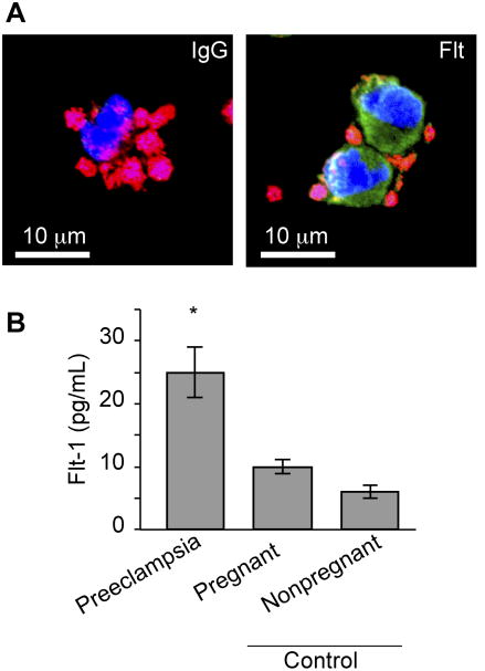
Figure 1. Flt-1 expression is increased in PMAs from women with preeclampsia.
(A) PMAs present in freshly-isolated mononuclear cells from women with preeclampsia were stained with WGA (red), which stains sialic acids in platelets and mononuclear cells and TOPRO-3 (blue), which stains nuclei. In parallel, an antibody specific for the N-terminus of Flt-1 (green) or its IgG control was used to identify Flt-1 in monocytes. (B) Freshly-isolated monocytes (including PMAs) were immediately lysed and total (soluble and membrane-bound) Flt-1 levels were determined by ELISA. The bars in this graph represent the mean ± SEM for all participants in each group, *p=0.003 compared to pregnant and nonpregnant controls.

