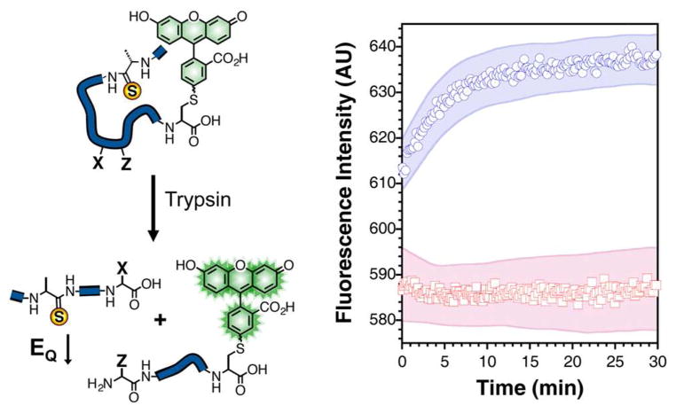
Figure 4.
Protease Activity. Left: A cartoon illustrating the cleavage of a profluorescent peptide substrate by a protease as described in the text. Right: Fluorescence of 1.4 μM A′AFKGψ peptide in the presence (blue circles) and absence (red squares) of 250 μg/mL trypsin in 67 mM sodium phosphate buffer, pH 7.6. Excitation was at 494 nm and emission was monitored at 522 nm. Shaded areas represent standard error as calculated from at least three independent trials.

