
Figure 5.
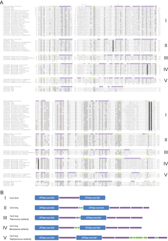
Multiple alignment of Csn2 subfamilies and comparison of their specific structural elements. (A) The multiple sequence alignment was constructed using the MUSCLE program for each Csn2 subfamily, separately. The alignments were then superimposed on the basis of conserved regions identified by HHPRED with some manual adjustment based on secondary structure predictions (see Supplementary Materials and Methods for details). The alignment with several ATPase sequences is based on Vector Alignment Search Tool (VAST) structural alignments with the structure of Csn2 of S. thermophilus (3ZTH) (17) used as a query (see Supplementary Materials and Methods for details). . The sequences are denoted by their GI numbers and species names. Secondary structure predictions and the secondary structure elements mapped to the respective crystal structures of the Csn2 long and short subfamilies are shown above the alignment for each Csn2 family. The positions of the first and last residues of the aligned region in the corresponding protein are indicated for each sequence. The numbers within the alignment represent poorly conserved inserts that are not shown. Secondary structure prediction is shown as follows: H indicates α-helix and E indicates extended conformation (β-strand). The positions strongly conserved in three families with a larger number of representatives are shown by reverse shading. The coloring is based on the 70% consensus built for a larger alignment (Supplementary Figure S7). Specific 90% consensus is also shown underneath the alignment for each family: ‘h’ indicates hydrophobic residues (WFYMLIVA), ‘c’ indicates charged residues (EDKRH) and ‘s’ indicates small residues (AGS). (B) Schematic representation of structures (actual and predicted) of five distinct Csn2 subfamilies. Cylindrical shape represents α-helix and arrow β-strand.