
Figure 6.
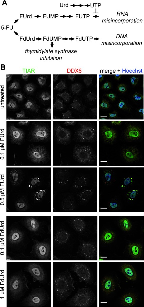
5-FU-induced SG assembly depends on RNA incorporation. (A) Schematic representation of the cellular 5-FU metabolism leading to incorporation of the different metabolites into RNA or DNA. (B) HeLa cells were treated with two concentrations of the 5-FU metabolites FUrd or FdUrd for 72 h. SG marker protein TIAR (green) and SG/P-body marker protein DDX6 (red) were visualized. Nuclei were stained with Hoechst. Scale bars represent 20 μm.