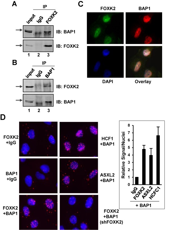
Figure 2.

Interactions between endogenous FOXK2 and BAP1. Reciprocal co-immunoprecipitation experiments using either FOXK2 (A) or BAP1 (B) antibodies for immunoprecipitation (IP) from U2OS cells. Co-precipitated endogenous BAP1 and FOXK2 were detected by immunoblotting (IB). Arrows represent the bands corresponding to each of the full-length proteins. (C) Immunofluorescence detection of endogenous FOXK2 (green) and BAP1 (red) and their co-localization in the nucleus [indicated by DAPI staining of DNA (blue)]. (D) Imaging and quantification of PLA signals generated by the indicated combinations of antibodies (IgG represents a non-specific antibody). DNA is stained using DAPI (blue) and the PLA signal is red. Quantitative analysis of PLA signals in the nucleus is shown on the right. The level of signals/nuclei in the control PLA sample (BAP1 and non-specific IgG antibodies) was set as 1.
