Figure 1.

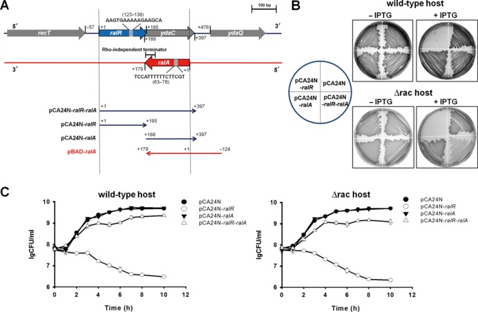
RalR is toxic and RalA reduces the toxicity of RalR. (A) The chromosomal region of ralR-ralA in E. coli is shown in the upper panel. ralR is shown by the blue arrow, ydaC is shown by the gray arrow, ralA is shown by the red arrow, and the ORFs in the neighborhood of ralR/ralA are shown as gray arrows. The direction and the specific region cloned into each of the plasmids used in this study are shown in the lower panel. The numbers indicate the position of the related nucleotides (position 1 on the sense strand indicates the first base A of the ralR ORF, and position 1 on the anti-sense strand indicates the first base G of RalA sRNA). (B) Cell growth in LB plates supplemented with chloramphenicol (30 μg/ml) with and without 0.5 mM IPTG for cells producing RalR and RalA in the wild-type host and in the Δrac host. (C) CFU test in LB medium supplemented with chloramphenicol (30 μg/ml) with 1 mM IPTG (added at OD600 0.1) in the wild-type host and in the Δrac host. Three independent cultures of each strain were evaluated.
