
Figure 1.
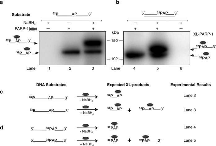
Cross-linking of purified PARP-1 to AP site-containing DNA. Phosphorimages of cross-linked PARP-1 to 32P-labeled AP site-containing DNA are shown in panels (a) and (b). Schematic representations of DNA substrates are shown above the phosphorimages. (a) Substrates were 5′-end 32P-labeled 34 bp DNA with a natural AP site or (b) 5′-end 32P-labeled dRP in a 34 bp DNA with a nick. Purified PARP-1 and 32P-labeled AP site DNA were incubated on ice without (-) or with (+) NaBH4 (see Materials and Methods). Lanes 1 and 6 in panels (a) and (b) represent DNA substrate incubations without PARP-1 as controls. The cross-linking products were analyzed by SDS-PAGE and followed by phosphorimaging. The migration positions of PARP-1 cross-linked to the full-length DNA strand (∼ 127 kDa), C3′ incised DNA strand (∼ 121 kDa), nicked DNA strand (∼ 122 kDa) and labeled dRP (∼ 116 kDa) are indicated. Panels (c) and (d) represent the substrates used in panels (a) and (b), respectively, and the expected cross-linked products under reaction conditions without (-) or with (+) NaBH4 are indicated. The symbol ( ) represents PARP-1, AP represents the AP site in the DNA, XL-PARP-1 means the cross-linked PARP-1 and 32P indicates the position of radiolabel on the DNA.