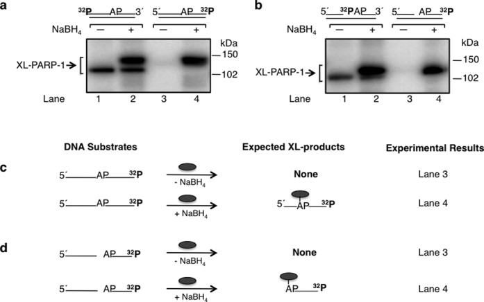
Figure 2.

Cross-linking of purified PARP-1 to AP site-containing DNA. Phosphorimages of cross-linked PARP-1 to 32P-labeled AP site-containing DNA are shown in panels (a) and (b). Schematic representations of DNA substrates are shown above the phosphorimages. (a) Substrates were 5′-end or 3′-end 32P-labeled 34 bp DNA with an AP site or (b) 5′-end 32P-labeled 34 bp DNA with an AP site in a nick (i.e. 5′-dRP) or 3′-end 32P-labeled 34 bp DNA with an AP site in a nick (i.e. 5′-dRP). The substrates in lanes 1 and 2 of panels (a) and (b) were the same as in panels (a) and (b) of Figure 1 and incubations were included for comparison. Purified PARP-1 and 32P-labeled DNA were incubated on ice without (-) or with (+) NaBH4 (see Materials and Methods). The cross-linking products were analyzed by SDS-PAGE and followed by phosphorimaging. The migration positions of the PARP-1 cross-linked with the full-length DNA strand, C3′ incised DNA strand, nicked DNA strand and labeled dRP are indicated. Panels (c) and (d) represent the substrates used in panels (a) and (b), respectively, and the expected labeled products under reaction conditions without (-) or with (+) NaBH4 are indicated; only one DNA strand is shown. The symbol ( ) represents PARP-1, AP represents the AP site in the DNA and 32P indicates the position of radiolabel on the DNA.
