Figure 1.
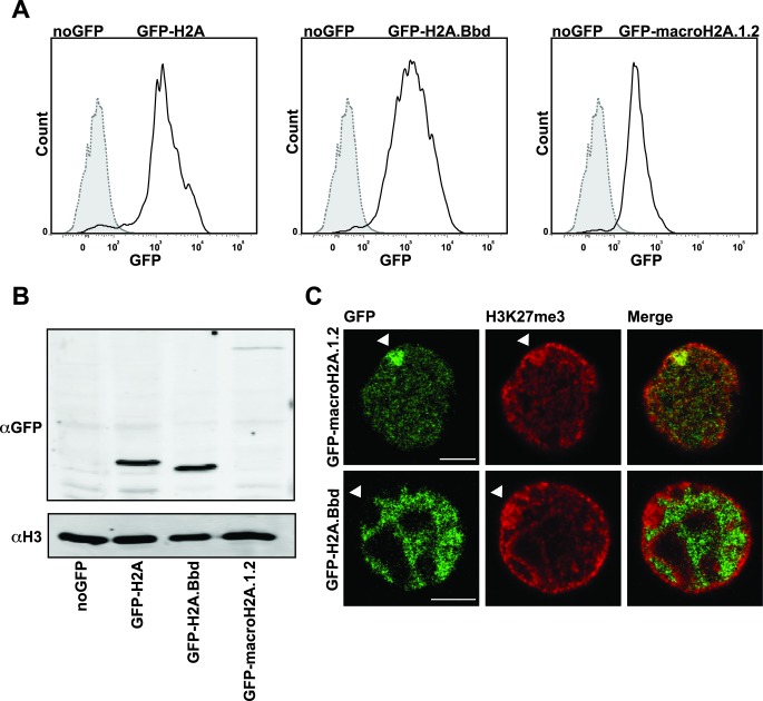
Female MEF single clones stably express the GFP-tagged histone variants. (A) FACS profile of female MEF cell populations derived from sorted single cell clones that express H2A.Bbd, macroH2A.1.2 and H2A tagged with GFP (solid line) in comparison to untransfected control female MEF cells (dotted and filled histogram). (B) Western blot analysis of GFP-tagged histone variant expression levels with antibody against GFP. Detection of histone H3 served as loading control. (C) Nuclear localization of the histone variants GFP-H2A.Bbd and -macroH2A.1.2 in female MEFs stained with H3K27me3 (red). Arrows indicate the Barr body. Scale bar: 5 μm.

