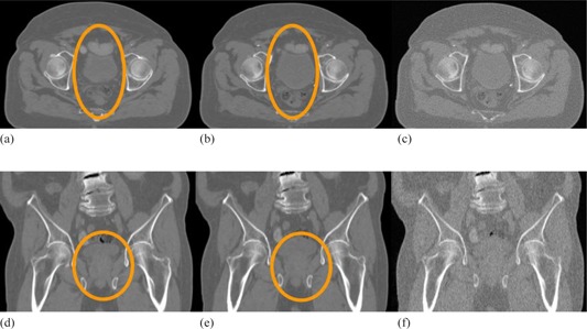
Figure 7.

Computational prostate phantom and simulated CBCT image sets. The axial cuts of (a) the original image set, (b) the deformed image set, and (c) the deformed image with simulated CBCT noise added. The subfigures (d)‐(f) are the coronal views of (a)‐(c), respectively.
