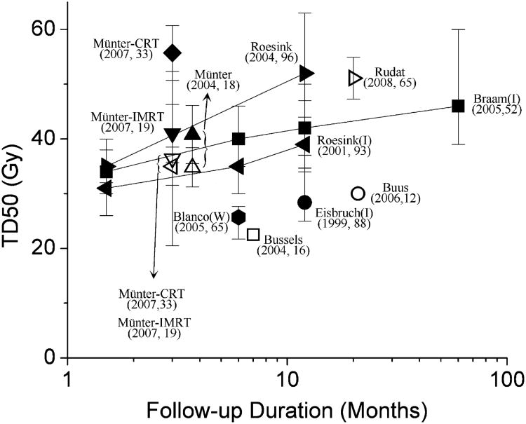
Fig. 3.
Reported tissue dose required for 50% response for loss of stimulated saliva flow after radiotherapy (RT) (2, 6, 10, 14–20) for single parotid gland. Endpoint considered in reports was salivary flow reduction to <25% (black symbols) or <50% (gray symbols) of pretreatment value. Tissue dose required for 50% response defined as dose at which 50% of patients developed complications. Error bars (if shown) indicate 95% confidence intervals; refer to original publications for exact meaning. 95% Confidence intervals for studies by Munter et al. (19, 20) were estimated from standard errors provided. Lines connect points from data sets with measurements taken at more than one interval after radiotherapy. Most studies used salivary gland scintigraphy. Some studies measured physical production (ipsilateral salivary flow or whole salivary flow; marked with “I” or “W”, respectively). Data from Buus et al. (2) (which did not include preradiotherapy assessments) derived by comparing different regions of parotid gland that had received different doses. Each label gives number of patients. Note, most imaging-derived endpoint data had greater values for tissue dose required for 50% response (TD50) than measured salivary data. CRT = conformal radiotherapy; IMRT = intensity-modulated radiotherapy.

