
Fig. 2.
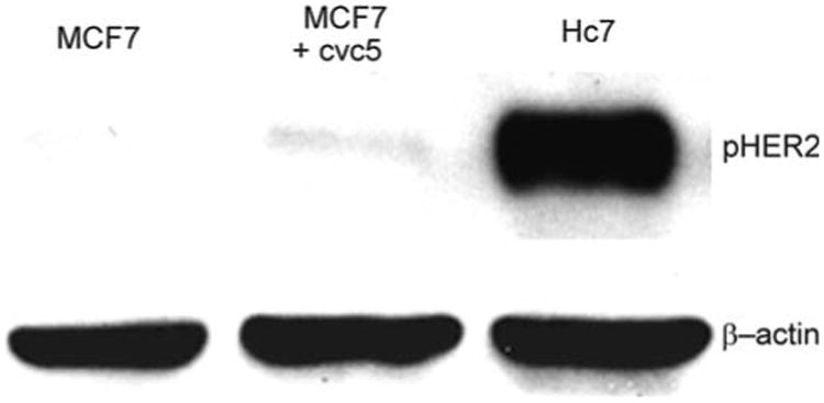
Western blots for HER2. Top row: Hc7 cell lysates show high expression of phospho-HER2 when compared to untransfected MCF7 cells or MCF7 cells transfected with the cvc5 vector, which lacks HER2 DNA. Bottom row: β-actin staining as loading control