
Fig. 5.
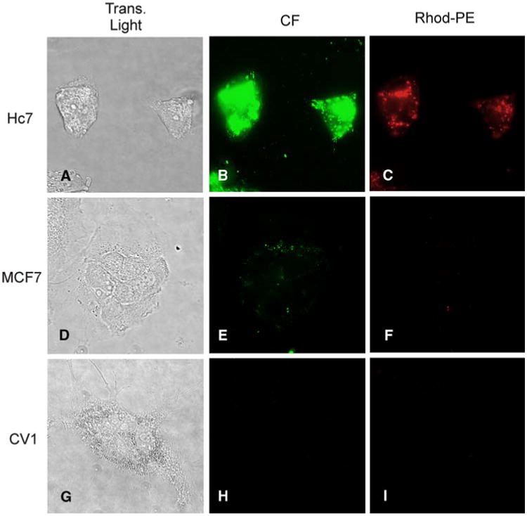
Incubation with anti-HER2 immunoliposomes containing Rhod-PE and encapsulating carboxyfluorescein (CF) generates bright intracellular signals in Hc7 cells, but not MCF7 or CV1 cells. Panels a, d, and g are transmitted light images; panels b, e, and h show fluorescence in the fluorescein channel; panels c, f, and i show fluorescence in the rhodamine channel. HER2-overexpressing Hc7 cells (a–c) avidly endocytose immunoliposomes, as evidenced by the intense red fluorescence of Rhod-PE (c). Endocytosis results in dequenching of CF to generate bright intracellular green fluorescence (b). MCF7 cells (d–f) express a physiological level of HER2 and endocytose fewer liposomes, giving rise to only feeble fluorescence. CV1 cells (g–i), which express no HER2, exhibit no fluorescence