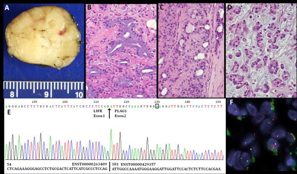
Figure 2.
LIFR-PLAG1 gene fusion in a soft tissue myoepithelial tumor (ME4). (A) Gross appearance of this well-circumscribed, yellow-tan, firm, hand subcutaneous lesion, arising in a 64 year-old female. The histologic spectrum of this tumor is quite broad, including: (B) ducts lined by columnar cells with abundant mucin-containing cytoplasm and basally-located nuclei; C. tubular structures lined by bland cuboidal epithelial cells; (D) Sheets of myoepithelial cells, with a distinctive rhabdoid or plasma-cell phenotype, showing densely eosinophilic cytoplasm and eccentric round nuclei. E.ABI sequence of 5’RACE product showed the presence of a fusion between LIFR exon 1 to PLAG1 exon 2; F. FISH showing a LIFR gene break-apart signal (telomeric probe , green; centromeric probe, orange).

