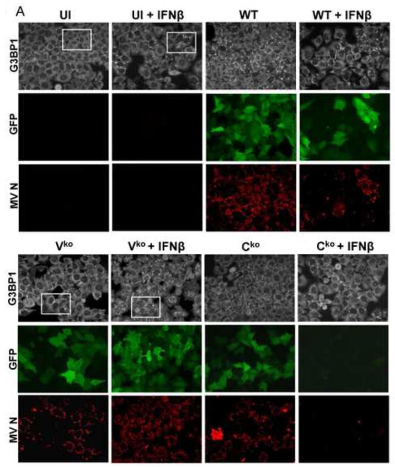
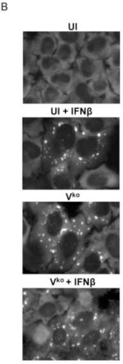
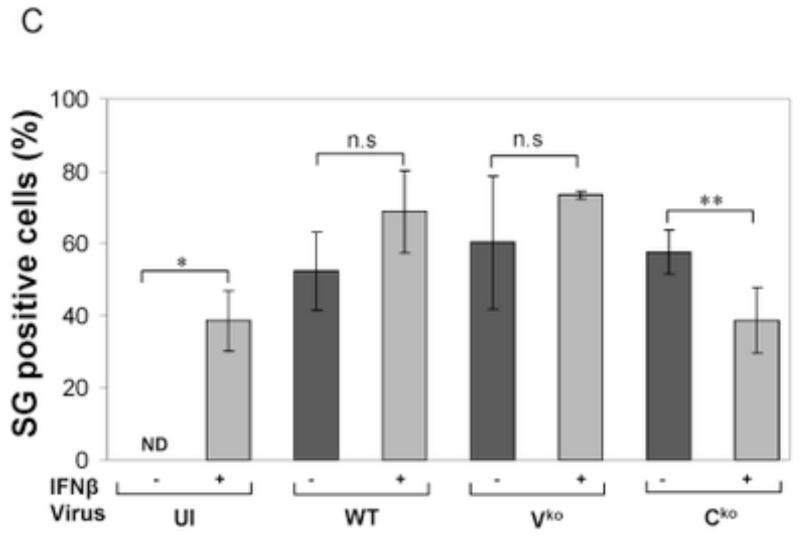
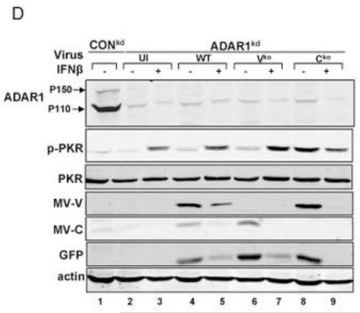
Figure 1. Stress granule formation is enhanced by interferon treatment of cells deficient in ADAR1.
(A) HeLa ADAR1kd cells were either left untreated or were treated with IFNβ (1000 U/mL) for 18 h. Cells then were either left uninfected (UI) or were infected with WT, Vko or Cko measles virus (MV) at a multiplicity of infection of 1 TCID50/cell. At 24 h after infection cells were analyzed by immunofluorescence microscopy as described under Materials and Methods using antibodies to G3BP1 (white) as a marker for SG formation and N (red) and GFP (green) for viral infection. (B) Magnification of the framed sections in panel A. (C) Quantification of SG-positive cells. Three representative wide-field 40× images were selected and a minimum of 100 cells were examined for the presence or absence of SG. Results are expressed as the percentage of infected cells that are positive for SG as described under the Methods section. Results are mean values and standard errors from three independent experiments. N.D., not detectable. Statistical significance determined by Students t test (two tailed, two sample with equal variance) comparing untreated cells to IFN treated cells. * P < 0.001, ** P < 0.05, n.s, not significant. (D) At 24 h after infection whole-cell extracts were prepared and analyzed by Western immunoblot assay using antibodies against ADAR1, phospho-Thr446 PKR (p-PKR), PKR, MV C protein, GFP, and actin.


