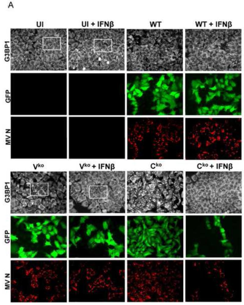
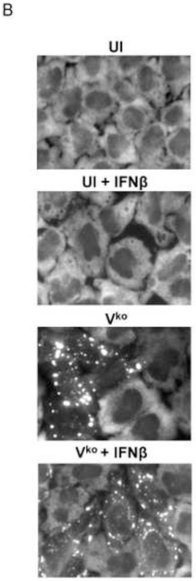
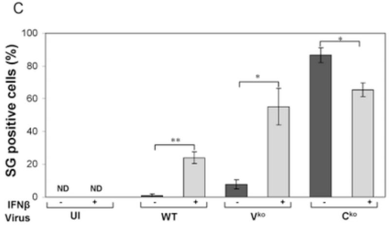
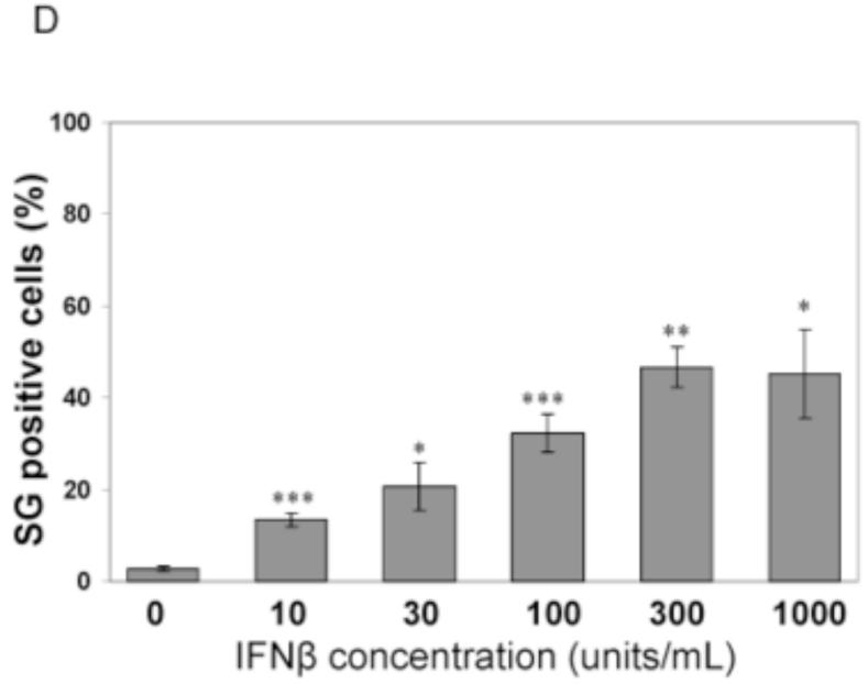
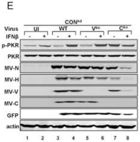
Figure 3. Interferon treatment enhances stress granule formation following infection with wild-type measles virus and mutant virus deficient for V-protein expression, but not with C-protein deficient mutant virus that already is an efficient inducer even in untreated cells.
(A) HeLa CONkd cells were either left untreated or treated with IFNβ (1000 U/mL) for 18 h. Cells then were either left uninfected (UI) or were infected with WT, Vko or Cko MV at a multiplicity of infection of 1 TICD50/cell. At 32 h after infection, cells were analyzed by immunofluorescence microscopy as described under Materials and Methods using antibodies to G3BP1 (white) as a marker for SG formation and N protein (red) and GFP (green) for viral infection. (B) Magnification of the framed sections in panel A. (C) Quantification of SG-positive cells as described for figure 1. Results are expressed as the percentage of infected cells that are positive for SG. Results are mean values and standard errors from three independent experiments. N.D., not detectable. Statistical significance determined by students t test (two tailed, two sample with equal variance) comparing untreated cells to IFN treated cells. * P < 0.005, ** P < 0.0005, n.s, not significant. (D) Quantification of SG positive cells when pretreated for 24 h with increasing concentrations of IFNβ prior to infection with Vko virus. Results are mean values and standard errors from three independent experiments. N.D.,not detectable. Statistical significance determined by Students t test (two tailed, two sample with equal variance) comparing untreated cells to IFN-treated cells * P<0.005, ** P<0.0005. (E) At 32 h after infection whole-cell extracts were prepared and analyzed by Western immunoblot assay using antibodies against phospho-Thr446 PKR (p-PKR), PKR, MV N, H, V and C proteins, GFP, and actin.

