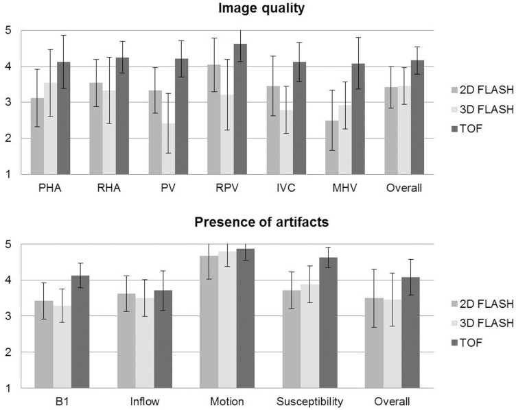
Figure 2. Qualitative analysis of image quality and presence of artifacts (5-point scales) for all analyzed liver vessel segments in all three sequences.
(PHA = proper hepatic artery, RHA = 2nd order branch of right hepatic artery, PV = main portal vein, RPV = 1st order branch of right portal vein, IVC = inferior vena cava, MHV = middle hepatic vein).

