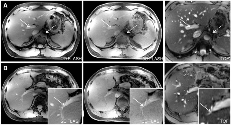
Figure 5. Depiction of the main portal vein (Figure A, arrows) and the right hepatic artery (Figure B, arrows) in all three sequences in one subject.
2D and 3D FLASH provided moderate to good image quality for both vessel segments with a slight advantage for 2D FLASH imaging due to a marginally higher contrast. TOF MRA was superior in the delineation of the liver arteries and veins from proximal to peripheral segments. Signal voids due to remaining B1-inhomogeneity were shifted out of the liver to a periaortal focus, causing minor impairment of vessel delineation (dashed arrow in Fig. A).

