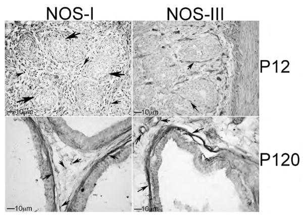
Figure 8.
Immunohistochemical analysis of NOS-I and NOS-III proteins in P12 and P120 prostate. At P12, NOS-I was identified in the mesenchyme in between the prostatic ducts while NOS-III was present in the basement membrane surrounding the ductal epithelium. In the adult prostate, NOS-I was abundant in neurons in the stroma between the ducts and in the basement membrane. At P12 and P120, NOS-III was restricted to the basement membrane surrounding the ductal epithelium and in the lining of blood vessels. 250× magnification. Small arrows indicate NOS-I and III staining. Large arrows indicate developing ducts of the prostate.

